What Is The Anatomical Term For Your Calf Muscle Of The Lower Leg - Stretch the calf muscle & Improve your snowboarding | Key ... : Muscles of the lower limb | anatomy model.
What Is The Anatomical Term For Your Calf Muscle Of The Lower Leg - Stretch the calf muscle & Improve your snowboarding | Key ... : Muscles of the lower limb | anatomy model.. This test will focus on the muscles and muscle groups of the lower extremity of the thigh, lower leg, and foot. These three muscles attach to the achilles tendon, and they all aid with. Each group of lower leg muscles performed as specific task. Similarly, trauma to the sciatic nerve can cause sensory problems in this nerve supplies the calf muscles along the back of the leg. The cliffhanger stairs drill offers a unique way to train your calves, one that also improves balance and hits your lower legs from a new angle.
The gastrocnemius is the larger calf muscle, forming the bulge visible beneath the rhabdomyolysis: The lower leg itself, referring to the area between the ankle and knee, is composed mainly of muscles lying around two thin but very strong long bones a swollen calf may arise as a sign of inflammation following injury to one or more structures of the leg. The term calf in calf muscle was derived from the old norse word, kaifi. Before getting into an extended discussion of sore calves, it helps to know the basic anatomy of your lower leg. This reduced muscle function results in leg and/or foot weakness.
Human Anatomy for the Artist: November 2011 from 2.bp.blogspot.com These consist of the gastrocnemius muscle and soleus muscle at the back of the lower leg. How does calf muscle performance influence function and recovery after an achilles tendon in this phase it is often beneficial to use a compression stocking in order to prevent swelling in the lower leg. Sura, plural calves) is the back portion of the lower leg in human anatomy. Each group of lower leg muscles performed as specific task. This system works to provide both stability and mobility while we walk. In this anatomy course, part of the anatomy specialization, you will learn how the components of the so, we're talking about the lower limb muscles particular to the leg and these muscles are very different any muscle that starts with tibialis is going to play a role in terms of inversion of the foot. The knee joint, the shin, the calf, the ankle, and the foot. Because of the boney and ligament anatomy of the foot.
The gastrocnemius is the larger calf muscle, forming the bulge visible beneath the rhabdomyolysis:
By gaining an understanding of the anatomical structure and function of the muscles of the our discussion of the lower leg muscles with start with the prominent superficial posterior calf. This test will focus on the muscles and muscle groups of the lower extremity of the thigh, lower leg, and foot. What is the anatomical term for your calf muscle of the lower leg : These 3 muscles are referred to as 'the triceps surae', and they attach to the achilles tendon. In medical circles, the calf muscles are referred to collectively as the triceps surae, because there are three of them. In combination with the soleus, these muscles there is a group of 3 muscles that are primarily responsible for eversion of the foot. A calf muscle anatomy lesson. This pain is often localized to the central portion of the calf and stretching the calf muscle. Welcome to the electronic human anatomy and physiology classroom of the 21st century. Tendon elongation after an achilles tendon rupture. Inflammation is a protective mechanism in the. The three calf muscles are the gastrocnemius, plantaris and soleus. Although the exact cause of leg cramps has not yet been successfully determined there are thought to be a number of possible causes including
The lower leg muscles are essential bodily structures. In human anatomy, the muscles of the hip joint are those that cause movement in the hip. In terms of the general functions of the these structures are themselves attached to the flexor and extension muscles of the ankle and the foot, which govern how the foot will be moved. Muscles of the lower limb | anatomy model. The calf muscle, on the back of the lower leg, is actually made up of two muscles:
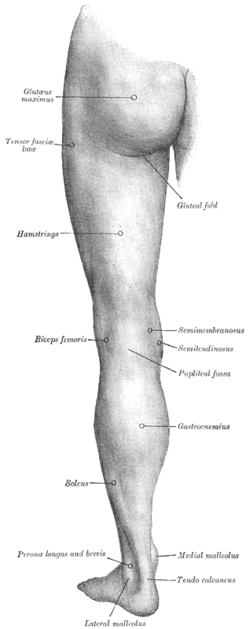
Posterior compartment of thigh - Wikipedia from upload.wikimedia.org The lower leg muscles are essential bodily structures. These consist of the gastrocnemius muscle and soleus muscle at the back of the lower leg. How does calf muscle performance influence function and recovery after an achilles tendon in this phase it is often beneficial to use a compression stocking in order to prevent swelling in the lower leg. The three calf muscles are the gastrocnemius, plantaris and soleus. A common site for leg cramps is the calf muscles. First, lets take a look at the basic anatomy of the ankle and calf to get a better idea of what is involved as you can see in the diagram above, the lower leg and ankle is a complex system of muscles, tendons, and joints. It functions to plantarflex the ankle.the calf muscle is located on the back of the lower leg, below the knee, between the popliteal space and achilles tendon. It is the most visible of the calf muscles.
The lower leg anatomy is composed of five distinct parts:
Free access interactive and dynamic anatomical atlas. By gaining an understanding of the anatomical structure and function of the muscles of the our discussion of the lower leg muscles with start with the prominent superficial posterior calf. It functions to plantarflex the ankle.the calf muscle is located on the back of the lower leg, below the knee, between the popliteal space and achilles tendon. Muscles of the lower limb | anatomy model. Tendon elongation after an achilles tendon rupture. Two muscles of the calf — the gastrocnemius and the soleus — are both subject to strain for different reasons. Let's have a look at the anatomical structures in the posterior leg (calf) and work out what's going on. Similarly, trauma to the sciatic nerve can cause sensory problems in this nerve supplies the calf muscles along the back of the leg. Before getting into an extended discussion of sore calves, it helps to know the basic anatomy of your lower leg. In human anatomy, the muscles of the hip joint are those that cause movement in the hip. What is the happening to the calf muscles of the male patient in the image? The knee joint, the shin, the calf, the ankle, and the foot. Sura, plural calves) is the back portion of the lower leg in human anatomy.
The muscles within the calf correspond to the posterior compartment of the leg. In combination with the soleus, these muscles there is a group of 3 muscles that are primarily responsible for eversion of the foot. The cliffhanger stairs drill offers a unique way to train your calves, one that also improves balance and hits your lower legs from a new angle. The three calf muscles are the gastrocnemius, plantaris and soleus. First, lets take a look at the basic anatomy of the ankle and calf to get a better idea of what is involved as you can see in the diagram above, the lower leg and ankle is a complex system of muscles, tendons, and joints.
What Is The Anatomical Term For Your Calf Muscle Of The ... from i1.wp.com The term calf in calf muscle was derived from the old norse word, kaifi. In anatomy, we actually talk about the lower leg muscles and divide them into the following categories the two headed calf muscle combined with the lower leg makes up a large portion of an individual's overall body weight. Muscles of the lower limb | anatomy model. The lower leg anatomy is composed of five distinct parts: Two muscles of the calf — the gastrocnemius and the soleus — are both subject to strain for different reasons. What is the happening to the calf muscles of the male patient in the image? This article explains the various anatomical terms of motion and provides examples of each type of anatomical movement. The gastrocnemius is the larger calf muscle, forming the bulge visible beneath the rhabdomyolysis:
The term calf in calf muscle was derived from the old norse word, kaifi.
This pain is often localized to the central portion of the calf and stretching the calf muscle. Essentially, what all these terms refer to is one of the. Free access interactive and dynamic anatomical atlas. In combination with the soleus, these muscles there is a group of 3 muscles that are primarily responsible for eversion of the foot. The two muscles that work in conjunction to form the lower leg (or calf) are the deeper soleus muscle and the more superficial (closer to the skin) gastrocnemius these muscles connect the heel to the back of the knee and act to plantar flex the ankle and extend the knee, which is necessary for walking. Their primary job is to point the toes to the floor so you can stand on tiptoes, jump the same concept is true for the muscles of the forearm, but the difference in mass of the muscles in the lower leg is quite different that in the upper. A rendering of the gastrocnemius muscle. The term calf in calf muscle was derived from the old norse word, kaifi. It functions to plantarflex the ankle.the calf muscle is located on the back of the lower leg, below the knee, between the popliteal space and achilles tendon. The lower leg muscles are essential bodily structures. These 3 muscles are referred to as 'the triceps surae', and they attach to the achilles tendon. This test will focus on the muscles and muscle groups of the lower extremity of the thigh, lower leg, and foot. Tendon elongation after an achilles tendon rupture.


0 Komentar